What is Trisomy?
Trisomy comes from the latin word "tri" which means three. "Somy" refers to an autosome, a type of chromosome (but not one of the sex chromosomes) which contains the genetic makeup of a person. Put very simply a trisomy is a chromosomal anomaly that is characterised by the presence of an extra chromosome in the cells of a person's (or plant or animal's) body.
When a Trisomy occurs it means that there are three identical chromosomes in a persons cell instead of the normal two.
Trisomy 18 (Edward's Syndrome) means that there are three 18th chromosomes,
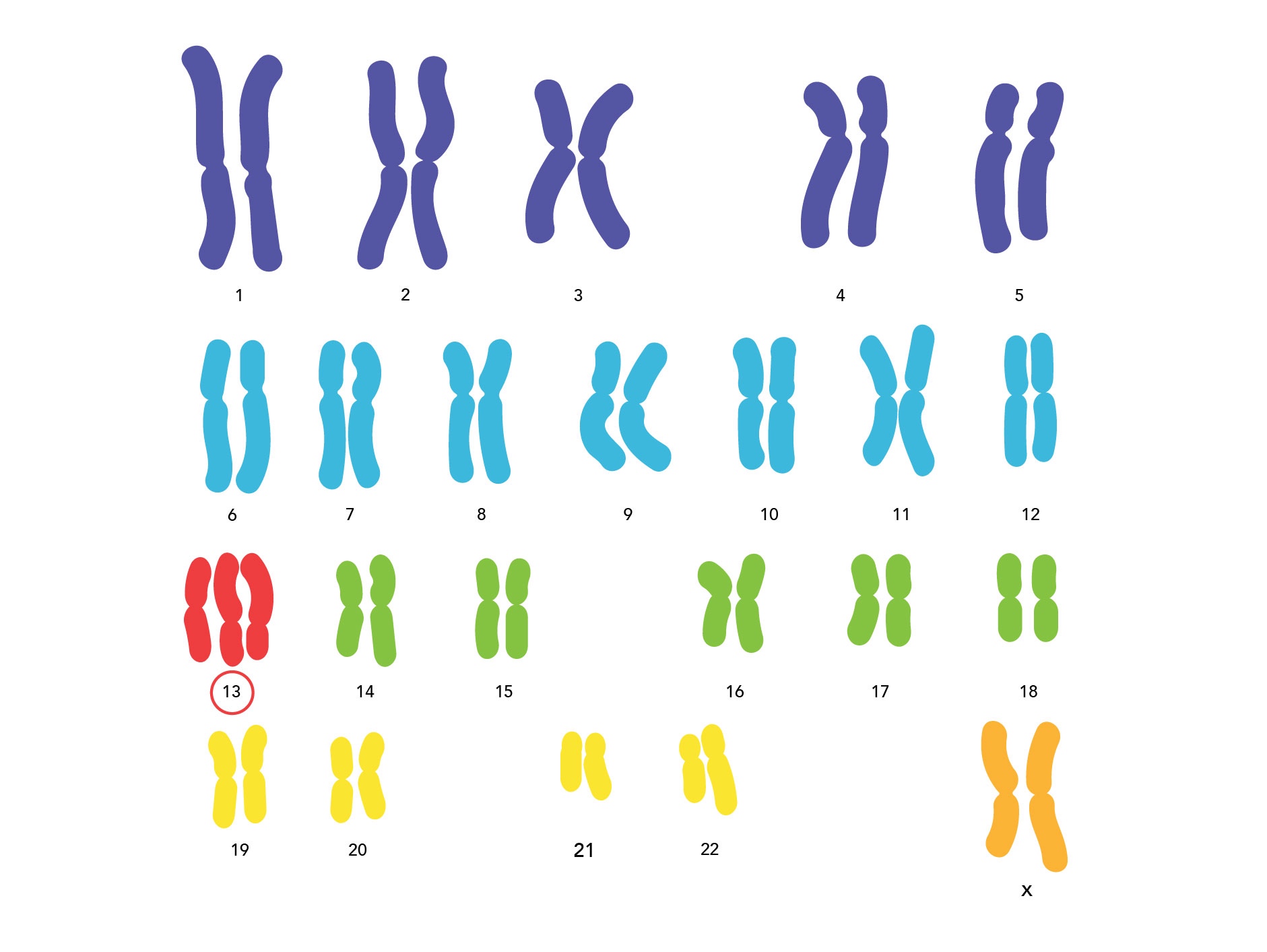
Trisomy 13 (Patau's Syndrome) means that there are three 13th chromosomes,
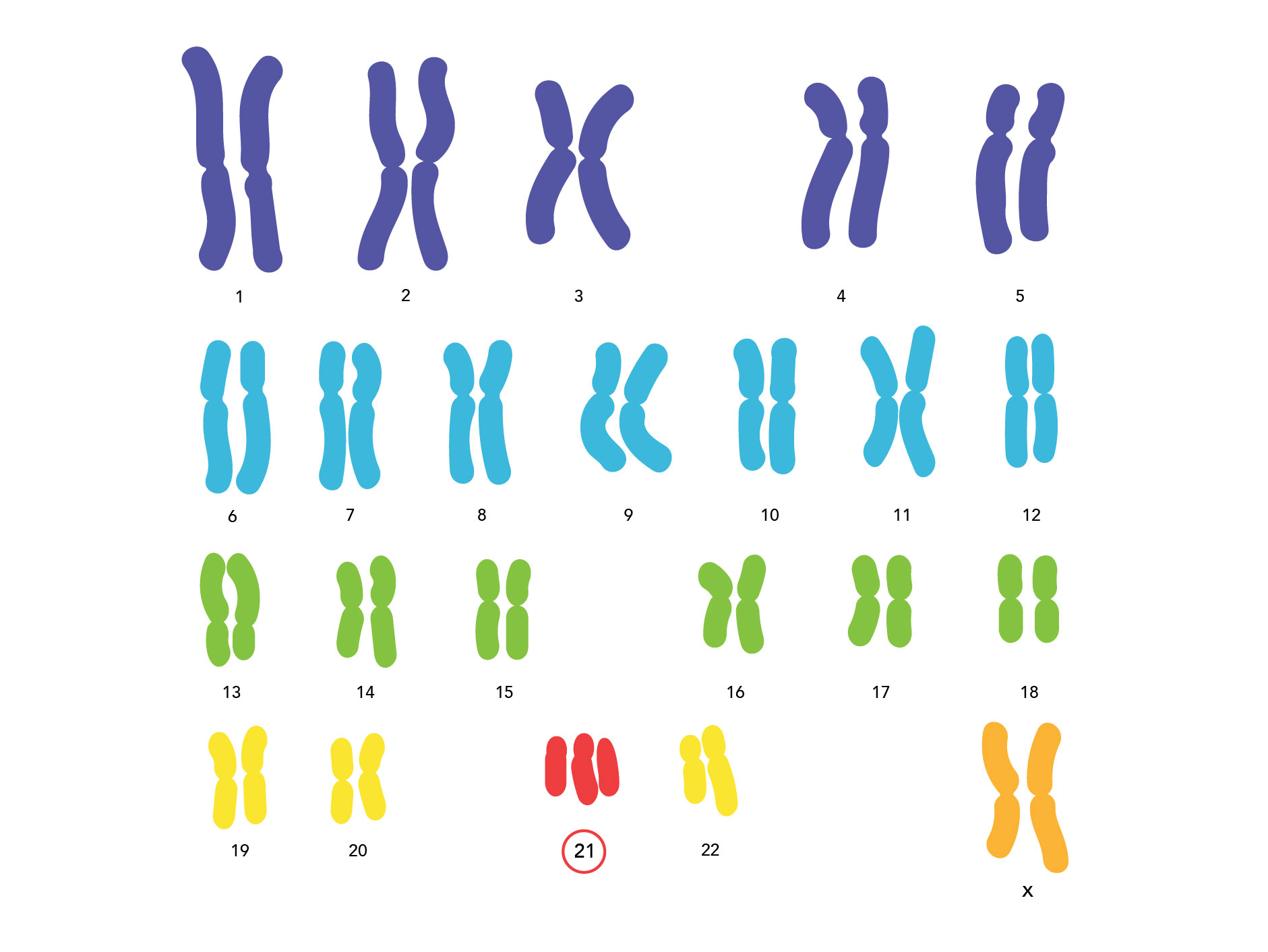
Trisomy 21 (Down Syndrome) means that there are three 21st chromosomes and so on.
A Trisomy can occur with any of the chromosomes. So you can have trisomy 1, trisomy 2, 3, 4, 5 and so on up to 22.
Chromosomes are tiny thread like structures which are contained in the nucleus of every human cell. In general every human cell contains 22 pairs of chromosomes that are known as autosomes and two sex chromosomes that are labelled X or Y.
It is these chromosomes which contain the genetic blueprint of life and which cause every person to be a unique individual.
Under normal circumstances everyone has a total of 46 chromosomes in each of their cells (including the sex chromosomes), a trisomy occurs where there are 47 chromosomes instead of the usual 46.
Each cell also contains a pair of sex chromosomes. In a male this combination is an X and a Y chromosome (XY). And in a femeale there are two X chromosomes (XX). One X chromosome is inherited from the mother and the other chromosome, be it an X or a Y, is inherited from the father. It is this chromosome, either an X or a Y, inherited from the father, which determines the sex of the baby.
Together the 22 pairs of autosomes and the two sex chromosomes make up a total of 46 chromosomes.

Standard human karyotype.
When we refer to a trisomy we are normally referring to the situation where every cell in the body contains the extra chromosome. Sometimes, in an effort to distinguish between other types of trisomy this is referred to as "full" trisomy.
A partial trisomy occurs when there is part of an extra chromosome, as opposed to a whole extra chromosome. Normally this is attached to another chromosome and is often (but not always) the result of one of the parents being the carriers of a balanced translocation. The effects of a partial trisomy on an individual vary from person to person depending on exactly how much of the extra chromosome that is duplicated.
Mosaic trisomy is where not every cell contains the extra chromosomal material. Some cells have the extra chromosome (47) and some cells have the usual compliment (46). The effects of mosaicism vary depending on how many cells are effected (the percentage) and where in the individuals body those cells are (the distribution).
This is a general overview of chromosomal anomalies as they relate to a trisomy. There are many other kinds including deletion syndromes, inversions, balanced translocations, Robertsonian translocations and more.
A trisomy can occur with any of the chromosomes, including the sex chromosomes. However the most commonly known trisomies are:
Trisomy 21 (Down Syndrome)
Trisomy 18 (Edward's Syndrome)
Trisomy 13 (Patau's Syndrome)
Trisomy of the sex chromosomes is often viewed differently from a trisomy of the autosomes because the genetic material contained on the sex chromosomes is very different to that of the autosomes.
Through research and the development of staining techniques in the 1950's, scientists were for the first time able to view the human chromosome. Although they appear disorganised within the cell, scientists have been able to identify them and so have numbered them from 1 to 22 in order of size. When this is done it is called a karyotype.
This information compiled and researched by Karen Schuler for SOFT Australia.

How are Trisomy conditions diagnosed?
Most cases of Trisomy are diagnosed prenatally in Australia. Regardless of whether the diagnosis is made prenatally (before birth) or postnatally (after birth) the process is the same. A sample of the baby’s DNA is extracted from a blood sample or other bodily cells or tissue and is cultured to examine a picture of the chromosomes called a karyotype.
A karyotype is simply a picture of a person’s chromosomes. In order to get this picture, the chromosomes are isolated, stained, and examined under the microscope. Most often, this is done using the chromosomes in the white blood cells. A picture of the chromosomes is taken through the microscope. A visible extra chromosome confirms a Trisomy diagnosis.

Prenatal Testing
Screening tests determine if the baby has an increased risk of having a particular problem such as Down syndrome T21, Edward’s Syndrome T18 or a neural tube defect. They are not diagnostic and an increased risk result does not mean the baby will definitely be affected.
Prenatal screening tests include:
Ultrasound
Early pregnancy (first trimester) screening: nuchal translucency ultrasound together with testing of the mother’s blood. The blood screen measures two pregnancy related hormones: hCG (human chorionic gonadotropin) and PAPP-A.
Pregnancy associated plasma protein A (PAPP–A) is a hormone that is produced by the placenta in pregnancy. Low levels of PAPP–A can be associated with Down's Syndrome (an extra chromosome 21), Edward's (extra chromosome 18) and Patau's syndrome (extra chromosome 13).
The ultrasound evaluation measures nuchal translucency (fluid beneath the skin behind baby's neck). This non-invasive procedure combines the results from the blood tests and the ultrasound, along with the mother's age, to determine risk factors.
Second trimester screening: testing the mother’s blood (maternal serum screening)
Diagnostic tests determine if the baby has, or will develop after birth, a genetic condition. Sampling procedures to obtain cells for chromosome analysis or specific genetic tests are invasive.
Postnatal Testing
Fluorescence in situ hybridization (FISH) is a test that "maps" the genetic material in human cells, including specific genes or portions of genes.
FISH is a method that can be used to detect small deletions and duplications that are not visible using microscope analysis. It can also be used to detect how many chromosomes of a certain type are present in each cell and to confirm rearrangements that are suspected after microscope analysis. FISH looks specifically at the one specific area of a chromosome only. It uses a very small chemical that glows brightly when it detects the specific region on a chromosome. A scientist uses a special microscope to look at the chromosome and see how many bright spots are present. When a person has a deletion, only one bright spot can be seen instead of two (one on each chromosome). When a person has a duplication, three bright spots can be seen instead of just two.
Why has FISH been offered for your child?
FISH is often performed alongside standard microscope analysis if your child has characteristics that are strongly suggestive of a particular deletion syndrome or another syndrome for which a FISH test is available. Your geneticist may request microscope analysis and FISH testing together or may request FISH testing if microscope analysis gives a normal result.
If after reading this information you have any questions we are more than happy to attempt to answer them. Just remember when undertaking any of these tests you do have choices, this is your pregnancy, your baby.
This information has been compiled by Tracey Pass on behalf of SOFT Australia with thanks to Trisomy 18 foundation, Babycentre.com, rarechromo.org and Wikipedia.
Helpful Links
ARCAN
We are a group of parents, who aim to help other parents and families. We have all walked the rare chromosome path and we are at different stages in our journey. With our experience and support we want to help other families who are also riding along this rare chromosome path.
SOFT USA
SOFT USA has been established for over 28 years and have many excellent resources available to parents
International Trisomy Alliance
9TIPS support for Trisomy 9
Heartfelt
Heartfelt is a volunteer organisation of professional photographers from all over Australia dedicated to giving the gift of photographic memories to families that have experienced stillbirths, premature births, or have children with serious and terminal illnesses.